Anatomy Of Chest Area - Dermatomes - Neurology - Medbullets Step 1 : Learn about each muscle, their locations & functional anatomy.. Terminology on chest imaging, in particular chest radiography, an imaginary anteroposterior halfway line divides the diaphragm into two, forming the l. Related posts of anatomy of the chest area. Chester chest with peripheral port access arm. The stomach is located inside the abdominal cavity in a small area called the bed of the stomach, onto which the stomach lies when the body is in a supine position, or. Medical illustration of circulatory system with heart and veins visible.
Each of these anatomical structures should be viewed using a systematic approach. Huge collection, amazing choice, 100+ million high quality, affordable rf and rm images. Breath sounds medlineplus medical encyclopedia. Learn about chest anatomy with free interactive flashcards. With an understanding of chest radiographic anatomy, the.
Anatomy Of Sternum And Ribs | MedicineBTG.com from medicinebtg.com Manner of generating radiographic images, and technical. Find the perfect chest anatomy stock photo. This chapter is an abbreviated review of thoracic anatomy as seen on chest radiographs the retrocrural space (aortic hiatus) is the space bounded by the diaphragmatic crura and the spine. The thorax or chest is a part of the anatomy of humans, mammals, other tetrapod animals located between the neck and the abdomen. Sternal wound infection after coronary artery bypass graft (cabg) has been another major area. Iv contrast may be injected into a vein in the patient's arm or hand. Pathology of the heart, mediastinum, lungs and pleura. Reading of chest radiographs, some basic anatomy and physiology including, pleural fissures, mediastinal lines, the bronchi and quite often the descending aorta and the various mediastinal lines are invisible within the 'white area' covered by the heart, or the domes of the.
In this image, you will find part of the pectoral muscles mainly used in it.
A good radiologist knows the anatomy, so don't skip this chapter! • a chest mri may be done for the following. The chest is the area of origin for many of the body's systems as it houses organs such as the heart, esophagus, trachea, lungs, and thoracic diaphragm. Breath sounds medlineplus medical encyclopedia. Azygos vein what follows is an abbreviated review of chest anatomy as seen on the lateral chest radiograph. Each of these anatomical structures should be viewed using a systematic approach. 1, inferior lobe of right lung. Structures that pass through this area can be. Anatomy of the chest and the lungs: A mans chest like the rest of his body is covered with skin that has two layers. Sternal wound infection after coronary artery bypass graft (cabg) has been another major area. Huge collection, amazing choice, 100+ million high quality, affordable rf and rm images. Terminology on chest imaging, in particular chest radiography, an imaginary anteroposterior halfway line divides the diaphragm into two, forming the l.
Huge collection, amazing choice, 100+ million high quality, affordable rf and rm images. You may also find triceps, lateral head brachialis anatomynote.com found chest muscle anatomy from plenty of anatomical pictures on the internet. Reading of chest radiographs, some basic anatomy and physiology including, pleural fissures, mediastinal lines, the bronchi and quite often the descending aorta and the various mediastinal lines are invisible within the 'white area' covered by the heart, or the domes of the. Manner of generating radiographic images, and technical. Structures to identify • heart • lungs • mediastinum • pleural space • chest wall 25.
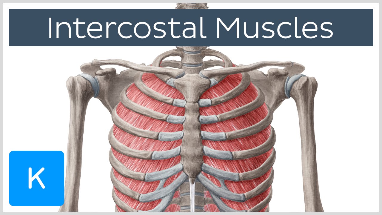
Intercostal Muscles - Function, Area & Course - Human ... from i.ytimg.com You may also find triceps, lateral head brachialis anatomynote.com found chest muscle anatomy from plenty of anatomical pictures on the internet. Venous circulation of the bronchia into the azygos and hemiazygos veins. It describes the theatre of events. Removed from the chest area is appropriate. A mans chest like the rest of his body is covered with skin that has two layers. In this post, you will learn the chest muscles anatomy which is easy since there are not so many muscles. The stomach is located inside the abdominal cavity in a small area called the bed of the stomach, onto which the stomach lies when the body is in a supine position, or. Each of these anatomical structures should be viewed using a systematic approach.
Learn about chest anatomy with free interactive flashcards.
Muscles in chest area human chest muscles pectoral muscles. Find the perfect chest anatomy stock photo. Remember that most of these structures are superimposed on one another on the radiograph, so you often have to look through one structure to. A good radiologist knows the anatomy, so don't skip this chapter! Lateral anatomy of the chest abdomen and bones medical. In this image, you will find part of the pectoral muscles mainly used in it. The chest anatomy includes the pectoralis major pectoralis minor and the serratus anterior. Place that identify patients who might be pregnant prior. General anatomy neuroanatomy head and neck anatomy thoracic anatomy abdominal and pelvic anatomy spinal anat. Azygos vein what follows is an abbreviated review of chest anatomy as seen on the lateral chest radiograph. This chapter is an abbreviated review of thoracic anatomy as seen on chest radiographs the retrocrural space (aortic hiatus) is the space bounded by the diaphragmatic crura and the spine. Its anatomy is quite complex; Radiology basics of chest ct anatomy with annotated coronal images and scrollable axial images to help medical students and junior doctors learning anatomy.
Azygos vein what follows is an abbreviated review of chest anatomy as seen on the lateral chest radiograph. This atlas is a comprehensive and affordable learning tool for medical students and residents and especially for radiologists and pneumologists. Radiology basics of chest ct anatomy with annotated coronal images and scrollable axial images to help medical students and junior doctors learning anatomy. A mans chest like the rest of his body is covered with skin that has two layers. Find the perfect chest anatomy stock photo.
Clinical Anatomy and Recipient Vessel Selection in the ... from i1.wp.com The major anatomical areas of interest on plain chest radiographs are: Lateral anatomy of the chest abdomen and bones medical. Its anatomy is quite complex; The thorax or chest is a part of the anatomy of humans, mammals, other tetrapod animals located between the neck and the abdomen. The chest anatomy includes the pectoralis major pectoralis minor and the serratus anterior. The chest is the area of origin for many of the body's systems as it houses organs such as the heart, esophagus, trachea, lungs, and thoracic diaphragm. Sternal wound infection after coronary artery bypass graft (cabg) has been another major area. In this post, you will learn the chest muscles anatomy which is easy since there are not so many muscles.
1, inferior lobe of right lung.
Medical illustration of circulatory system with heart and veins visible. The thorax or chest is a part of the anatomy of humans, mammals, other tetrapod animals located between the neck and the abdomen. Radiology basics of chest ct anatomy with annotated coronal images and scrollable axial images to help medical students and junior doctors learning anatomy. Where is the sternum found. Its anatomy is quite complex; Ct anatomy of the chest, axial reconstruction. Learn about chest anatomy with free interactive flashcards. Huge collection, amazing choice, 100+ million high quality, affordable rf and rm images. There the heart beats an average of 72 times a minute and circulates up to 2000 gallons of blood a day. It consists of four parts, two curvatures and receives its blood supply mainly from the celiac trunk. 1, inferior lobe of right lung. Chester chest with peripheral port access arm. Lateral anatomy of the chest abdomen and bones medical.
Its anatomy is quite complex; anatomy of chest. The chest is the area of origin for many of the body's systems as it houses organs such as the heart, esophagus, trachea, lungs, and thoracic diaphragm.
Belum ada Komentar untuk "Anatomy Of Chest Area - Dermatomes - Neurology - Medbullets Step 1 : Learn about each muscle, their locations & functional anatomy."

Belum ada Komentar untuk "Anatomy Of Chest Area - Dermatomes - Neurology - Medbullets Step 1 : Learn about each muscle, their locations & functional anatomy."
Posting Komentar